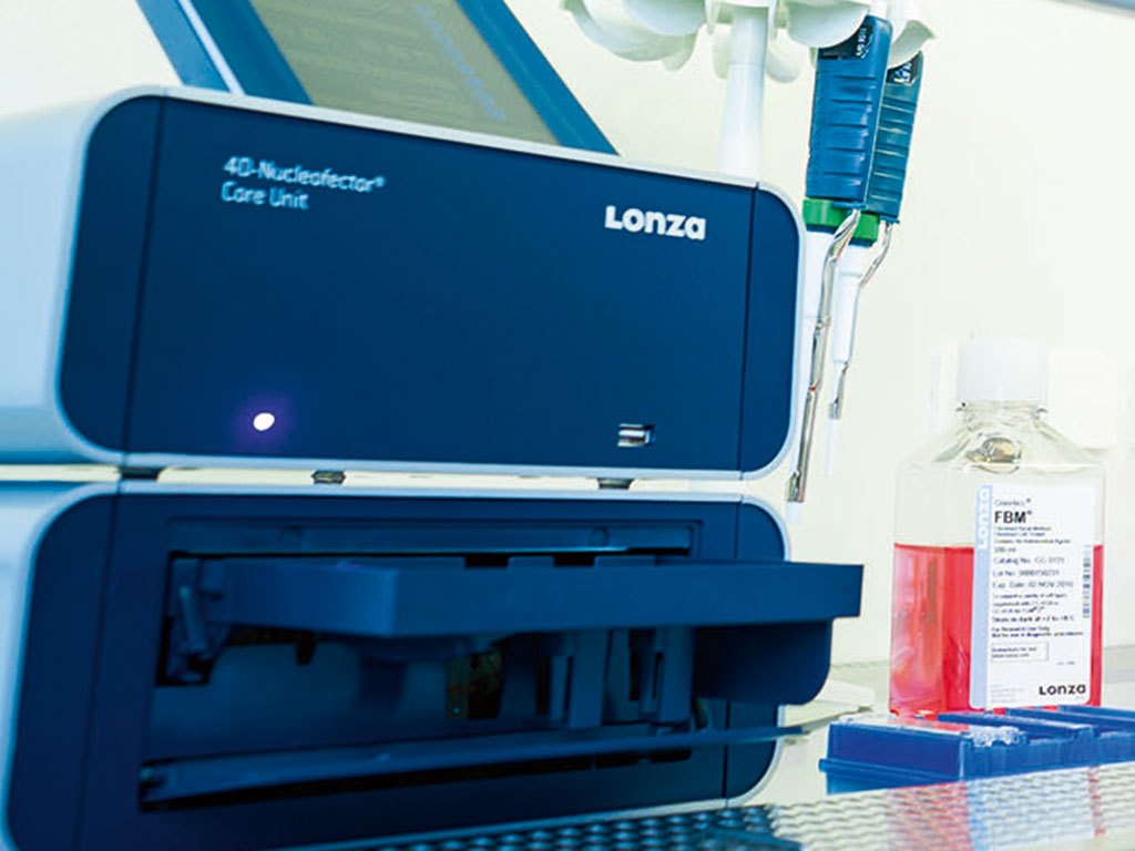
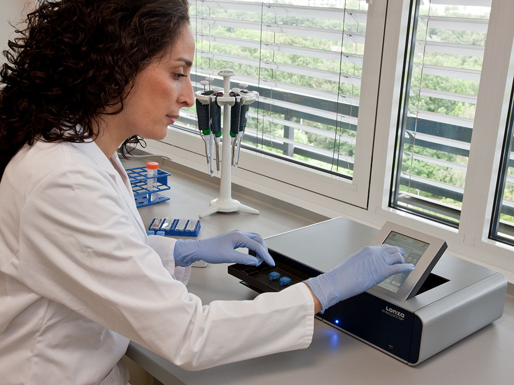
Did you know that SAECs (small airway epithelial cells) are a crucial model for target identification and validation in drug discovery? During our virtual event about CRISPR in Drug Discovery Anna Dickson and Alessia Serrano (Senior Research Scientists at AstraZeneca) were two of our fantastic speakers.*
Recently, Dickson et al. published a scalable and automated CRISPR screening workflow for SAECs combined with a disease-specific endpoint assay in SLAS Discovery.
As primary cells such as SAECs are hard to transfect cells, CRISPR/Cas9 gene editing is often performed by ribonucleoprotein (RNP) nucleofection. An established and robust CRISPR screen is not only a powerful tool for drug target identification, but also for the target validation associated with disease phenotypes of primary cell types. The detailed screening protocol utilizes the advantages of both; a disease-relevant immortalised cell line can be utilized for a larger scale screen (e.g. genome wide) and then the top hits can be carried forward into a smaller, targeted validation screen using disease-relevant primary cells such as SAECs.
As it is impractical to assess the efficiency of editing of each individual gene in a screening, it is critical to include editing controls and genes which drive or reduce the desired endpoint phenotype to ensure confidence in the whole automation workflow. It is also crucial to add a neutral editing control for normalisation instead of unedited cells as CRISPR gene editing itself can affect cell health or growth.
We put together a short summary of the SAEC CRISPR screen workflow with the use of RNPs (for further details see Dickson et al.), 384-well Nucleofector® System and P3 Primary Cell 384-well Nucleofector® Kit.
Workflow
First step: Cell culture of SAECs (thawing and expansion for 8 days to achieve 30 Mio. cells)
Second step: Preparation of RNP plate and complex formation (2h)
Third step: CRISPR editing of SAECs via 384-well Nucleofector® System(4h)
Fourth step: Cell handling post Nucleofection® (2h)
Fifth step: Dispensing the edited cells into assay plates
Sixth step: Endpoint assay and analysis (4-5 days after CRISPR gene editing)
The publication also gives relevant information regarding critical steps, troubleshooting (e.g. errors due to bubbles) and can be easily adapted to other cell types.
If you are interested or planning CRISPR screening experiments, we highly recommend to read through this great publication.
We thank Anna Dickson and Alessia Serrano for their brilliant talks at our virtual event CRISPR in Drug Discovery. Of note, also other speakers spoke about setting up CRISPR screens – watch it on demand.
Written by
Camilla Scientific
Support Specialist
References:
Dickson A, Mullooly N, Serrano A, Escudero-Ibarz L, Wiggins C, Gianni D. Highly scalable arrayed CRISPR mediated gene silencing in primary lung small airway epithelial cells. SLAS Discov. 2023 Mar;28(2):29-35
Important note: The user bears the sole responsibility for determining the existence of any third party rights, as well as obtaining any necessary licenses, related to performing the screening workflow, including using CRISPR/Cas9.
*The statements made by each of the presenters does not necessarily represent the opinions of his or her employer.