Cryopreserved ampule of Human Umbilical Vein Endothelial Cells (HUVEC), from a single donor, in EGMTM-2 containing ≥ 500,000 cells

Cryopreserved ampule of Human Umbilical Vein Endothelial Cells (HUVEC) from pooled donors, cultured in EGMTM-2, pre-screened for Angiogenesis and containing ≥ 500,000 cells
Cryopreserved ampule of Human Umbilical Vein Endothelial Cells (HUVEC), from pooled donors, in EGMTM-2 and containing ≥ 500,000 cells
Cryopreserved ampule of Human Human Umbilical Vein Endothelial Cells (HUVEC) from a single donor containing ≥ 500,000 cells
Cryopreserved ampule of Human Umbilical Vein Endothelial Cells (HUVEC) from pooled donors, containing ≥ 500,000 cells
Cryopreserved ampule of Human Umbilical Vein Endothelial Cells (HUVEC) without VEGF, from a single donor, cultured in EGMTM Plus and containing ≥ 500,000 cells
Basal media required for growth of Endothelial cells, 500 mL (supplements and growth factors sold separately)
-
TechSheet - Umbilical Vein Endothelial Cell SystemsTechnical information sheet for CloneticsTM HUVEC cells
-
Instructions for Use - HUVEC Culturing and Angiogenesis Inhibition Assay using Sartorius Incucyte®Protocol for Angiogenesis assay using HUVEC cells
-
Tube Formation Assay with Primary HUVECsIn this study, we have optimized this assay using Lonza's Primary HUVECs and EGM®-2 Media
-
Expanded HUVECs for High-throughput ScreeningHUVECs are a good primary endothelial cell type for many in vitro studies and also for several high-throughput screening applications. Consequently, Lonza offers, HUVEC-XL. These cells are expanded to passage-3 from pooled P1-HUVECs and packaged at 10 million cells per ampoule.
-
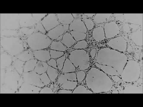 Tube Formation Assay with HUVECHuman Umbilical Vein Endothelial Cells seeded on BME and the formation of endothelial tubes were monitored live with the CytoSMART™ Lux 10X System, the previous version of the CytoSMART™ 2 System. Time-lapse video was recorded for 22 hours at a frame interval of 1 frame every 5 minutes.
 -
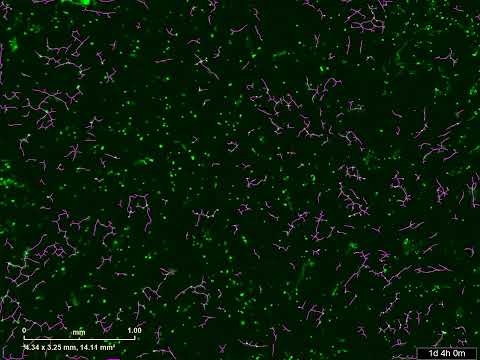
 HUVEC-Fibroblast Co-Culture - AngiogenesisTime-Lapse Video of Human Umbilical Vein Endothelial Cell Angiogenic Tube Formation in Co-Culture with Dermal Fibroblasts. Primary Human Umbilical Vein Endothelial Cells (HUVECs) were co-cultured with primary Human Normal Dermal Fibroblast cells at a ratio of 1:3 HUVECs: fibroblasts and a total cell density of 40,000 cells/well in a 96-well plate. Co-cultures were maintained for 5 days with medium changes every 2-3 days in EBMTM-2 medium supplemented with a custom concentration of VEGF (313 pM) to optimize angiogenic tube formation in this setting. Cells were stained with CellTracker® Dye from ThermoFisher. Purple lines indicate angiogenic tube formation detected by the live cell imaging software. At each frame, the software quantifies angiogenesis by counting the number of network branch points (i.e., the number of branches in each network of tubes) per mm2 and the mean network length (i.e., the average of the total network length of tubes per mm2).
 -
 HUVEC-Fibroblast Co-Culture – Angiogenesis Inhibition
 -
Automated Cell Signaling Assays Using Primary HUVECsLonza's Primary HUVECs are an excellent cell type to measure the effects of pharmacologically-important compounds on vial signaling pathways, and they provide increased data relevancy compared to cell lines.
-
Lonza Extends Industry-Standard HUVEC Product LineLonza, the market leader in primary cells, has expanded its human primary cell portfolio with an improved version of Human Umbilical Vein Endothelial Cells (HUVEC) cultured in the absence of additive vascular endothelial growth factor (VEGF).
-
Instructions - HUVEC-XL™ Pooled Cell SystemInstructions for culturing Clonetics™ HIVEC-XL™Pooled Cells