Discontinuation notice: This product has been discontinued as of 31 Dec, 2019. Please visit Cytosmart Technologies BV for purchase and continued support.
Added to Your Shopping Cart
Certificate of Analysis
Are you looking for...
You might be interested in...
Type in Product name, Keyword or Catalog number to see suggestions.
Need help? Please contact us
Save to list
Save to list
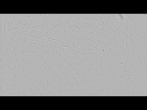
Cell Culture Monitoring with CytoSMART™ System
The composition of culture media, the nature of culture vessels and culture matrices, and the subculture procedure are known to have a significant impact on cell culture and should be standardized wherever possible. Defining standard operating procedures (SOPs) for cell culture helps to reduce the variables affecting cell culture performance and assay results. Subculture details, such as culture confluency at the time of splitting, seeding density and total number of passages, should be precisely defined.
However, even a standardized cell culture experiences variation as a biological system. For example, primary cell systems such as patient derived materials may vary widely in proliferation behavior. Additionally, cell culture performance is highly user dependent and requires a certain degree of expertise to achieve consistency in cell cultures. Regardless of these challenges, standardized cell culture is of great importance as the condition of a cell culture may have a significant influence on the downstream experiment results obtained using the cultured cells.
Microscopes, small enough to fit inside a cell culture incubator and at the same time offering live cell imaging and time-lapse recording functionalities can help monitoring cell cultures. Automatic cell confluency determination provided by some microscopes can reduce operator dependent variations in cell culture handling. Unfortunately, most microscopes are not suited to be operated inside an incubator, or they are too big to be used for or too valuable to be blocked by cell culture monitoring applications.
1
of
Unbiased Determination of Cell Culture Confluency
1
of

In addition, automatic alerts can be set so that the researcher receives an email notification once the cell culture has reached a chosen confluency, thus offering a very simple yet effective tool to easily standardize cell culture.
Time-lapse Microscopy: Video Recording of Cell Cultures
1
of

Lonza News with Leigh Eastwood show what is really happening in your incubator and how suspicious cell conduct can be avoided!

The CytoSMART™ System. How it works.
Setting up a CytoSMART™ Live Recording requires minimal training and you’ll be up and running in minutes. Learn how easy-to-use a CytoSMART™ System is.Growth Curve Recording with the CytoSMART™ System
1
of
This video of human astrocytes, recorded with the CytoSMART™ 2 System, shows an untypical proliferation rate of NHAs. Corrective actions need to be taken.
Growth Curve of NHA Culture
The growth curve of the same Normal Human Astrocyte culture recorded with the CytoSMART™ 2 System indicates a flat growth curve revealing a nontypical proliferation behaviour of NHAs. Corrective actions need to be taken.Standardization of Subculturing Monolayer Cells
Subculturing monolayer cultures is a routine task in every cell culture lab. To minimize the impact on cell culture quality and assay outcome, the procedure should be standardized wherever possible. While some variables,such as culture medium, dissociation agent or cell culture vessel are easy to mitigate, the time point of subculture and the dissociation itself remain user dependent.
The CyoSMARTTM System with its growth curve recording and automatic alert function enables you to always subculture your cells at the same cell density and avoid subjective determination of confluency. Cells should be in log phase (exponential growth phase) when subcultured, ideally at 70 to 80% confluency.
The detachment of the cells from the cell culture vessel is one of the most important steps in the subculturing process and is, unfortunately hard to standardize. When using an enzymatic dissociation agent, the temperature and the freshness have an important impact on the enzymatic activity of the enzyme and thus on the detachment. Ideally the enzyme should be aliquoted and for each subculture a fresh aliquot should be used. Depending on how strongly cells attach to their culture vessel surface, the dissociation can be performed at room temperature or in an incubator. In any case, it is important to monitor the dissociation under a microscope to avoid damage to the cells. This observation is easily done when cells are incubated with the enzyme at room temperature. Use of the CytoSMARTTM System means that close observation is possible even in the incubator. Put your cell culture with the enzymatic agent on the CytoSMARTTM System in the incubator and observe the cells lifting from the culture vessel from the tablet. The ideal time point to stop the enzymatic activity by adding serum containing media, PBS/BSA, Trypsin inhibitor, or Trypsin Neutralization Solution is when the cells are rounded up and only a few cells start to float.
Hypoxic Culture of Cells Monitored with CytoSMART™ System
Hypoxic Culture in Stem Cell Applications
Hypoxic culture conditions are thought to support stem cell performance in general. The use of physiologically relevant oxygen levels in stem cell culture is beneficial, for example, for the generation of induced pluripotent stem cells (iPSC) and in mesenchymal stem cell (MSC) culture. iPSCs have the ability to self-renew and generate any cell type in the human body. Therefore, they have the potential to produce an infinite quantity of cells for different applications, such as regenerative medicine, disease modeling and drug development. Many efforts have been made to find ways to facilitate the reprogramming process as well as the maintenance of iPSCs. Culturing iPSCs in undisturbed hypoxic conditions has been found to be one of the options that can elicit significant improvements to stem cell culture.
Image of hMSCs Cultured under Hypoxic Conditions
The image of Human Mesenchymal Stem Cells cultured in 5 % O2 was taken with the CytoSMARTTM Lux 10X Device. (Image courtesy of A. Henn, BioSpherix)
Enhanced iPSC generation, reduced spontaneous differentiation, and enhanced clonality of human embryonic stem cells have been demonstrated to be a result of hypoxia-induced stabilization of transcription factors of the Hypoxia-inducible factor (HIF) family.
The use of human mesenchymal stem cells (hMSCs) as a possible therapeutic tool in regenerative medicine has been widely assessed. Poor growth kinetics, early senescence, and genetic instability during in vitro expansion are among the major challenges for MSC-based regenerative therapies. By applying physiological oxygen levels to the cell culture, major improvements have been achieved, such as modulated angiogenic potential of MSCs, reduced senescence and increased proliferation levels1.
Furthermore, the quantity and quality of MSCs secreted bioactive molecules (called MSC secretome) change under hypoxic conditions. The number of different proteins expressed in hMSCs cultured under hypoxic conditions is much higher (see graph to the left), and secreted proteins that are identical between hypoxic and room air oxygen conditions show significantly different expression levels. These findings support why the MSC secretome is being considered a critical element for therapeutic efficacy of hMSC.
Analysis of human umbilical cord Wharton Jelly-derived hMSCs conditioned medium (hWJ-MSC CM): More proteins were identified in the hWJ-MSC CM collected from hypoxic conditions (166 proteins) when compared to the normal atmospheric oxygen conditions hWJ-MSC CM (104 proteins), in which 81 were common to the two conditions1.
Remote Cell Culture Monitoring under GMP Conditions
Now that cell therapy and immunotherapy approaches are making their way from the bench into patients, more and more cells are cultured under good manufacturing conditions (GMP) in dedicated GMP suites. Each time a scientist enters the suite, a complex gowning routine has to be followed including hair net, shoe covers, gloves, mask, coverall, boot covers, googles, and sterile gloves. Under these circumstances, live cell monitoring is costly and quite time consuming; there is no option for researchers to go take a quick glance at their cells.
As a cloud-based system, the CytoSMARTTM System for live cell imaging and monitoring enables you to view your cell culture from any browser-capable system, such as a computer, laptop, smartphone or personal tablet device. Moreover, you can set automatic alerts for cell confluence - meaning you only need to physically tend to your cells once they have reached a certain confluency.
Although the CytoSMARTTM System is not compliant to GMP regulations (e.g. no 21 CFR Part 11 compliance), using it can reduce the number of times cell cultures need to be manually observed.
The same advantage applies to biosafety level (BSL) 3 & 4 laboratories, labs that allow diagnostic work and research on pathogens which can cause severe or fatal diseases. Working in these BSL 3 or 4 labs is extremely cumbersome and costly. In addition to the gowning, researchers also need to disinfect themselves when exiting the lab. Thus, using the remote monitoring feature of the CytpoSMARTTM System not only reduces costs (costs of gowning equipment) but also increases the safety of the employees.
Cell Counting with the CytoSMART™ System
The new CytoSMARTTM System has been developed for live cell imaging and monitoring. In addition, the CytoSMARTTM System offers a quick and easy-to-use cell counting feature using a standard hemocytometer. Within seconds, the CytoSMARTTM App determines the exact number of cells in your culture. If cells are stained with Trypan blue, the number of live cells is detected.