-
 CompareeLearning: Troubleshooting the BETCatalog #: LAL-EL-6This module explains different types of endotoxin test failures for gel clot and kinetic LAL assays, guides you through possible causes and suggests strategies to help avoid these issues.Sales unit: each
 -
CompareeLearning: Understanding the BETCatalog #: LAL-EL-2This introductory module introduces assay mechanisms, the basic assay requirements, the need for endotoxin controls and how Limulus Amebocyte Lysate (LAL) is made.Sales unit: each
-
CompareHUVEC – Human Umbilical Vein Endothelial Cells, Single Donor, in EGM™-2Catalog #: C2517A
Cryopreserved ampule of Human Umbilical Vein Endothelial Cells (HUVEC), from a single donor, in EGMTM-2 containing ≥ 500,000 cells
Sales unit: each -
CompareXTREME® CHO Feed Liquid – Chemically Defined and Protein-free, 1 LCatalog #: BE02-056Q
XTREME® CHO Feed Liquid - CD, Protein free, Liquid, 1L
Sales unit: each -
CompareHuman Mesenchymal Stem Cell (hMSC) Chondrogenic Differentiation Medium BulletKit®Catalog #: PT-3003
Culture system containing hMSC Chondrogenic Basal Medium (PT-3925) and hMSC Chondrogenic SingleQuots® Kit Supplement (PT-4121) required for hMSC chondrogenic differentiation
Sales unit: kit -
ComparehMSC Human Mesenchymal Stem Cell Chondrogenic Differentiation Basal MediumCatalog #: PT-3925hMSC Chondrogenic Differentiation Basal Medium, 185 mLSales unit: each
-
ComparehMSC Human Mesenchymal Stem Cell Chondrogenic Differentiation Medium SingleQuots® Supplements and Growth FactorsCatalog #: PT-4121
hMSC Chondrogenic Differentiation SingleQuots® Kit
Sales unit: each -
CompareHUVEC – Human Umbilical Vein Endothelial Cells, Pooled, in EGM™-2, Prescreened for AngiogenesisCatalog #: C2519AS
Cryopreserved ampule of Human Umbilical Vein Endothelial Cells (HUVEC) from pooled donors, cultured in EGMTM-2, pre-screened for Angiogenesis and containing ≥ 500,000 cells
Sales unit: Ampule -
CompareHUVEC - Human Umbilical Vein Endothelial Cells without VEGF, Single Donor, in EGM™-PlusCatalog #: CC-2935
Cryopreserved ampule of Human Umbilical Vein Endothelial Cells (HUVEC) without VEGF, from a single donor, cultured in EGMTM Plus and containing ≥ 500,000 cells
Sales unit: Ampule -
CompareBovine Brain Extract (BBE), 5 mlCatalog #: CC-4098
Bovine Brain Extract contains endothelial cell proliferation growth factors, 9 mg/mL, 5 mL
Sales unit: each
-
Instructions - Cryopreserved Kupffer CellsInstructions for Thawing and Plating of Human Cryopreserved Kupffer Cells
-
NPCs Hepatic Non-Parenchymal CellsThese additional liver cell types provide a complete building set supporting complex cell culture modeling. Applications include developing more physiologically relevant in vitro models for complex liver diseases and toxicity testing of new drugs and chemicals.
-
The Importance of Cell-subtype Selection in Creation of a 3D Liver Fibrosis ModelData sheet detailing 3D liver fibrosis model in collaboration with Visikol
-
Inventory - Non-Parenchymal CellsListing of available non-parenchymal cells inventory; updated monthly March 2019 NPC Inventory
-
Primary cells and other tools for in vitro ADME-ToxCD-LI019_ADME-Tox_Product_listing_sec.pdf
-
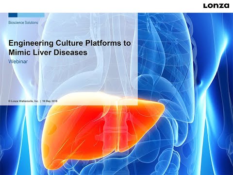 Engineering Culture Platforms to Mimic Liver DiseasesLearn from Dr. Salman Khetani, University of Illinois at Chicago, how multiple liver cell-types including hepatocytes, Kupffer, stellate, and endothelial cells can be engineered together to model human liver disease phenotypes in culture.
 -
TechSheet – Dulbecco’s Phosphate Buffered Saline (DPBS)Product use of Dulbeccos Phosphate Buffered Saline (DPBS).
-
A Novel In Vitro Liver Cell Culture Flow System Allowing Long-Term Metabolism and Hepatotoxicity Studies]Manuscript published in Applied In Vitro Toxicology journal describes improvement of CYP450 phenotype of primary cryopreserved hepatocytes using Quasi Vivo® System
-
Endothelial Cells Concentration Guide
-
 Live-cell imaging of CD34+ cells with Nanolive’s 3D Cell ExplorerHuman cord blood CD34+ stem/progenitor cells were cultured in X-VIVO™ 15 serum-free hematopoietic cell medium, in the presence of recombinant human thrombopoietin (25ng/mL), Rlt3 ligand (25ng/mL) and stem cell factor (13ng/mL). The culture was coated with fibronectin. Live cell imaging was performed with Nanolive’s 3D Cell Explorer for 15 hours (3 images/min during 15hours) and cell migration was captured.