Cryopreserved ampoule of Human Human Umbilical Vein Endothelial Cells (HUVEC) from a single donor containing ≥ 500,000 cells
Cryopreserved ampoule of Human Umbilical Vein Endothelial Cells (HUVEC) from pooled donors, containing ≥ 500,000 cells
Culture system containing EBM® Basal Medium (CC-3121) and EGM® Endothelial Cell Growth Medium SingleQuots® Supplements (CC-4133) required for growth of endothelial cells. (Pack of 6)
EGM®-Plus Endothelial SingleQuot® Kit
The Nebula® Absorbance Reader is a 96-well microplate reader that brings new and improved technology to the laboratory and is optimized to work with WinKQCL® Endotoxin Detection and Analysis Software and our traditional LAL assays such as the PYROGENT® 5000 Kinetic Turbidimetric Assay and the Kinetic-QCL® Kinetic Chromogenic Assay.
Cryopreserved ampule of Human Umbilical Vein Endothelial Cells (HUVEC), from pooled donors, in EGM®-2 Media and containing ≥ 500,000 cells
Cryopreserved ampoule of Human Umbilical Vein Endothelial Cells (HUVEC), from a single donor, in EGM®-2 Media, containing ≥ 500,000 cells
Culture system containing EBM®-2 Basal Medium (CC-3156) and EGM®-2 MV Microvascular Endothelial Cell Growth Medium SingleQuots® supplements (CC-4147) required for growth of Microvascular Endothelial Cells
Culture system containing EBM®-2 Basal Medium (CC-3156) and EGM®-2 SingleQuotsTM Supplements (CC-4176) required for growth of Endothelial Cells (Pack of 6).

-
Instructions - UltraCHO™ MediumHow to use ProCHO medium
-
Instructions – ProCHO Liquid mediaInstructions for use of ProCHO Media
-
Instructions ProCHO powder kitsInstructions for use of ProCHO Powder kits
-
ProCHO® Protein Expression Media
-
WinKQCL™ 5 Remote Client Installation InstructionsWinKQCL™ 5 client installation system requirements and configuration.
-
WinKQCL 5 Software - EnglishWe Analyze Endotoxin Data Every Day Version 5 Software Features Announcement of discontinued support for Windows SQL Server® 2000 and XP Operating System English Version
-
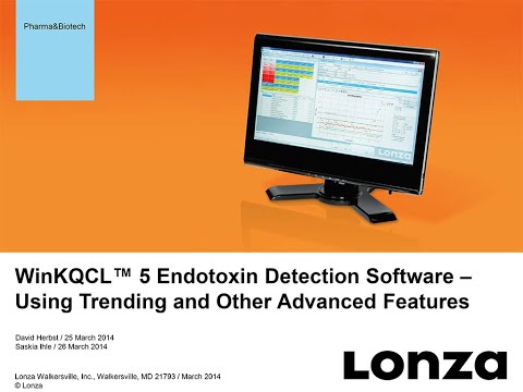 WinKQCL™ 5 Endotoxin Detection SoftwareLearn how to use the advanced features in Lonza's WinKQCL™ Endotoxin Detection Software to save time, reduce costs and ensure compliance to 21 CFR Part 11 and Annex 11.
 -
BE02-056Q CHO Xtreme™ Feed CD - InstructionsBE02-056Q
-
4D-Nucleofector® Protocol for MRC-5Optimized 384-well Nucleofector® and 4D-Nucleofector® Protocol for MRC-5
-
MRC-5 Transfection Protocol - 4D-Nucleofector™ DeviceInstructions of use for transfection of MRC-5 with the SE Cell Line 4D-Nucleofector™ X Kit