Added to Your Shopping Cart
Certificate of Analysis
Are you looking for...
You might be interested in...
Type in Product name, Keyword or Catalog number to see suggestions.
Looking for a Certificate of Analysis?
Have your catalog and lot numbers ready and click the link.
Find CoA
You searched for "kupffer cell"
Too many choices? Use the filter option on the left to narrow your product listing.
Compare
Human CD19+ B Cells (10 Million Cells, Negative Selection)
Catalog #: 4W-601
Human CD19+ B cells isolated from peripheral blood using negative immunomagnetic selection, cryopreserved, ≥10 million cells
Compare
Human CD19+ B Cells (10 Million Cells, Negative Selection)
Catalog #: 2W-601
Human CD19+ B cells isolated from peripheral blood using negative immunomagnetic selection, cryopreserved, ≥10 million cells
Compare
PrEC – Human Prostate Epithelial Cells
Catalog #: CC-2555
Cryopreserved ampule of Prostate Epithelial Cells (PrEC) containing ≥500,000 cells
Compare
hMSC - Human Mesenchymal Stem Cells, 750,000
Catalog #: PT-2501
Cryopreserved ampule of Human Mesenchymal Stem Cells (hMSCs) containing ≥750,000 cells
Compare
HprAD - Human Subcutaneous Preadipocyte Cells, 4 Million
Catalog #: PT-5001
Cryopreserved ampule of Human Subcutaneous Preadipocytes containing ≥4 million cells
Compare
Rat Brain Hippocampus Neurons
Catalog #: R-HI-501
Cryopreserved ampule of Rat Brain Hippocampus Neurons containing ≥ 1 million cells
Compare
HAEC – Human Aortic Endothelial Cells
Catalog #: CC-2535
Cryopreserved ampule of Human Aortic Endothelial Cells (HAEC) containing ≥ 500,000 cells
Compare
Human Liver-Derived Endothelial Cells P1
Catalog #: HLECP1
Low passage endothelial cells dervied from human liver tissue
Compare
NHDC - Human Dendritic Cells, 2.5 Million
Catalog #: CC-2701
Cryopreserved ampule of Normal Human Dendritic Cells (NHDC) containing ≥2.5 million cells
Compare
PyroCell® Monocyte Activation Test Human Serum Rapid System
Catalog #: 00296408
The PyroCell® Monocyte Activation Test – Human Serum (HS) Rapid System is an all-inclusive kit to conduct the Monocyte Activation Test.
Added to Your Shopping Cart
-
Instructions - UltraCHO™ MediumHow to use ProCHO medium
-
Instructions – ProCHO Liquid mediaInstructions for use of ProCHO Media
-
Instructions ProCHO powder kitsInstructions for use of ProCHO Powder kits
-
ProCHO® Protein Expression Media
-
WinKQCL™ 5 Remote Client Installation InstructionsWinKQCL™ 5 client installation system requirements and configuration.
-
WinKQCL 5 Software - EnglishWe Analyze Endotoxin Data Every Day Version 5 Software Features Announcement of discontinued support for Windows SQL Server® 2000 and XP Operating System English Version
-
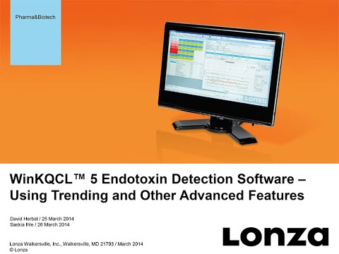 WinKQCL™ 5 Endotoxin Detection SoftwareLearn how to use the advanced features in Lonza's WinKQCL™ Endotoxin Detection Software to save time, reduce costs and ensure compliance to 21 CFR Part 11 and Annex 11.
 -
BE02-056Q CHO Xtreme™ Feed CD - InstructionsBE02-056Q
-
4D-Nucleofector® Protocol for MRC-5Optimized 384-well Nucleofector® and 4D-Nucleofector® Protocol for MRC-5
-
MRC-5 Transfection Protocol - 4D-Nucleofector™ DeviceInstructions of use for transfection of MRC-5 with the SE Cell Line 4D-Nucleofector™ X Kit
Product Availability by Store Location
Hours