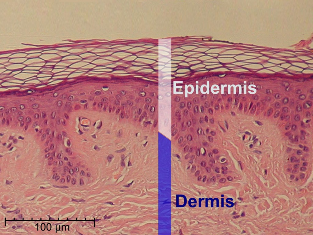
Normal Human Skin Tissue. Image courtesy of Wikimedia.
Dermal fibroblasts are cells within the dermis layer of skin which are responsible for generating connective tissue and allowing the skin to recover from injury.3 Using organelles (particularly the rough endoplasmic reticulum), dermal fibroblasts generate and maintain the connective tissue which unites separate cell layers. Furthermore, these dermal fibroblasts produce the protein molecules including laminin and fibronectin which comprise the extracellular matrix. By creating the extracellular matrix between the dermis and epidermis, fibroblasts allow the epithelial cells of the epidermis to affix the matrix, thereby allowing the epidermal cells to effectively join together to form the top layer of the skin.
Epidermal-dermal skin substitutes are currently the most utilized tissue-engineered skin model that closely resembles the structure of native human skin. The presence of both keratinocytes and fibroblasts within the epidermal-dermal skin substitutes leads to the production of a variety of growth factors and cytokines which expedite wound healing, highlighting the importance of epithelial-mesenchymal interactions. These epidermal-dermal skin substitutes have been utilized for wound healing assays, toxicological research, co-culture studies, tissue engineering research.